Fake Eyelashes and Mites: What Really Happens to Eye Health

Key facts
- Researchers at the National Pirogov Memorial Medical University in Vinnytsia, Ukraine, presented findings on eyelash extensions and mites at the 44th ESCR Congress
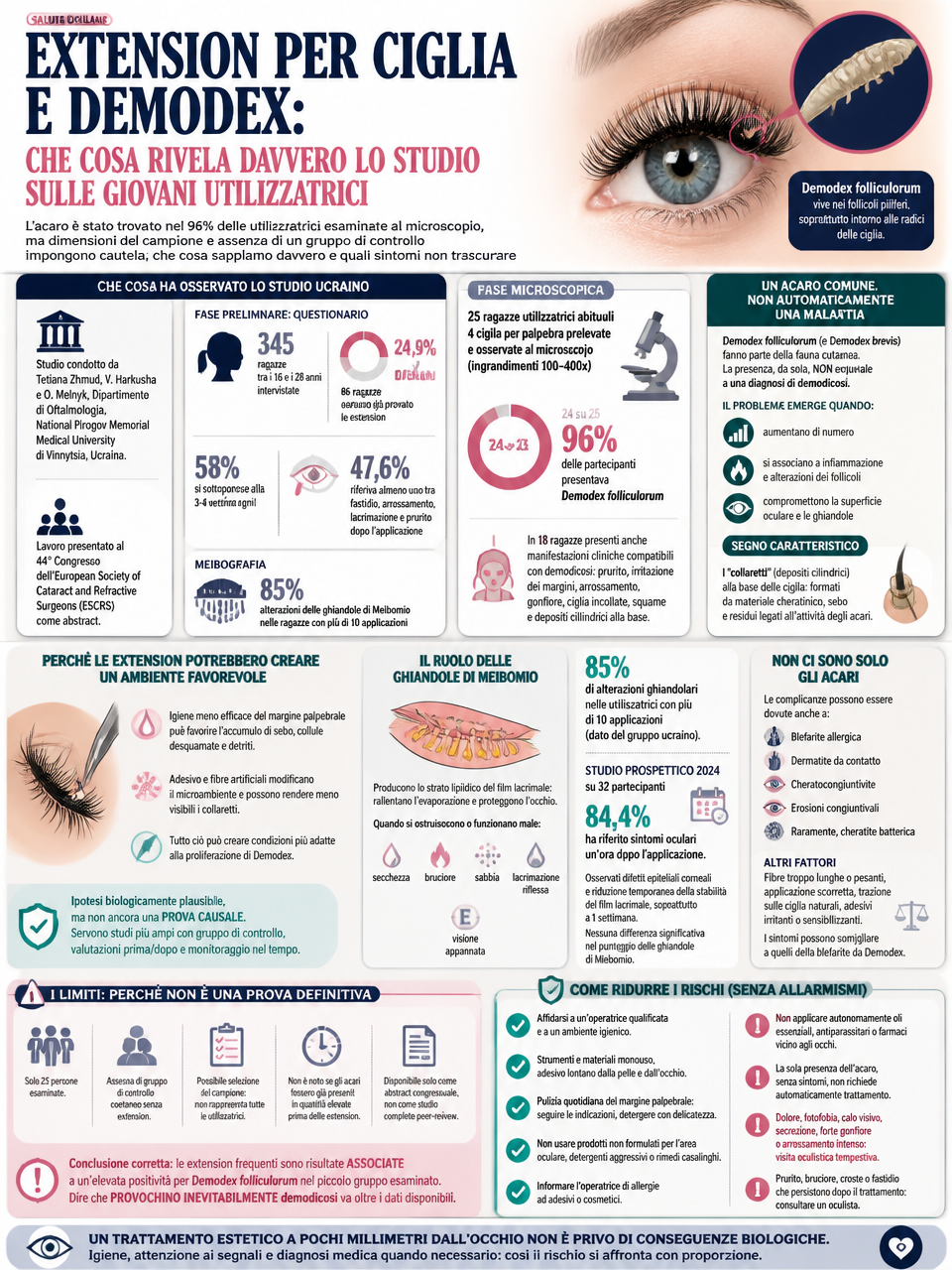
- In a small cohort of young extension users, researchers detected the Demodex folliculorum mite in 24 out of 25 cases
- A survey of 345 women aged 16 to 28 found that 24.9 percent had previously used eyelash extensions
- Among surveyed extension users, 58 percent reported undergoing the procedure every three to four weeks
- A 2024 prospective study involving 32 participants reported ocular symptoms in 84.4 percent of subjects one hour after application
At the base of the eyelashes, concealed within adhesive, sebum, and minute skin debris, conditions can emerge that favor the proliferation of a mite already native to the human face: Demodex folliculorum. In a small cohort of young, regular extension users, researchers detected the parasite in 24 out of 25 cases. While this finding is significant, it does not establish that artificial lashes directly cause infestations.
What the Ukrainian study observed
The study was carried out by Tetiana Zhmud, V. Harkusha and O. Melnyk from the Department of Ophthalmology at the National Pirogov Memorial Medical University in Vinnytsia, Ukraine. The findings were presented as a conference abstract at the 44th Congress of the European Society of Cataract and Refractive Surgeons (ESCRS).
In the initial phase, the researchers distributed a questionnaire to 345 women aged between 16 and 28. Of this group, 86, representing 24.9%, had previously used eyelash extensions. Among those who had used the product, 58% reported undergoing the procedure every three to four weeks, while 47.6% indicated experiencing at least one of the following symptoms after application: discomfort, redness, tearing or itching.
Meibography, an examination that allows for the visualization of the Meibomian glands, further revealed abnormalities in 85% of the participants who had undergone the procedure more than ten times. These preliminary data prompted the research team to investigate the potential presence of Demodex folliculorum among regular users.
The microscopic phase of the study involved 25 women who regularly used eyelash extensions. Four lashes were collected from each participant’s eyelid and subsequently examined under an optical microscope at magnifications ranging from 100 to 400 times. Mites were detected in 24 of the 25 participants, representing 96% of the small cohort analyzed. Eighteen of the women also exhibited clinical signs consistent with demodicosis, including itching, irritation of the eyelid margins, redness, swelling, lashes stuck together, and the presence of scales and cylindrical deposits at the base of the lashes.
While the 96% figure draws significant attention, it must be interpreted within its specific context. It does not indicate that 96% of all individuals wearing extensions will develop a disease, nor does it prove that extensions are the direct cause of mite proliferation. The statistic exclusively describes findings observed in a very narrow and selected subgroup of regular users.
A common mite, not automatically a disease
Demodex folliculorum is a microscopic organism that inhabits hair follicles, particularly in sebum-rich areas of the face. On the eyelids, it tends to cluster around the base of the eyelashes. A second species, Demodex brevis, penetrates more deeply into sebaceous units and the Meibomian glands.
The mere presence of these mites does not constitute a diagnosis of demodicosis. In many individuals, they are part of the normal skin flora and do not cause symptoms. Problems may arise when their numbers increase or when they are associated with inflammation, follicular changes, or ocular surface disorders. Scientific literature links clinically significant infestations to blepharitis, irritation, conjunctivitis, recurrent chalazia, and, in some cases, corneal issues; however, the pathogenic role can vary from person to person and must be assessed alongside clinical signs.
A distinctive indicator is the presence of so-called collarettes, cuffs, or cylindrical deposits encircling the base of the eyelashes. These structures consist of keratinous material, sebum, and debris linked to mite activity. In a study comparing patients with and without such deposits, Demodex was detected in every individual exhibiting widespread or sporadic cylindrical dandruff, whereas the prevalence was significantly lower among those lacking this sign. Consequently, examination of the eyelid margin, supplemented by microscopic analysis of the lashes when necessary, enables specialists to differentiate between mere colonization and a clinically significant condition.
Why false lashes may create a favorable environment
Eyelash extensions are bonded to natural lashes, either individually or in small clusters, using an adhesive applied near the hairline. This area houses the follicles and the openings of structures critical to maintaining the ocular surface’s homeostasis. To prevent compromising the bond, some users may reduce the frequency or thoroughness of their daily cleaning of the eyelid margin.
The researchers propose that inadequate hygiene and alterations to the skin’s microenvironment may facilitate the buildup of sebum, shed skin cells, and debris around the lash roots, creating conditions conducive to the proliferation of Demodex. Furthermore, the adhesive and synthetic fibers can obscure the telltale collarettes, potentially delaying the recognition of the issue.
The biological rationale behind this claim is sound, yet it does not constitute causal evidence. Establishing a definitive link would require broader investigations featuring a control group of peers who do not wear extensions, assessments conducted both prior to and following application, and longitudinal monitoring. Such studies would also need to account for other variables that can impact eyelid health, including cleaning methods and frequency, the type of adhesive used, the duration of wear, underlying conditions such as dermatitis or rosacea, contact lens usage, and any history of blepharitis.
The Role of the Meibomian Glands
The meibomian glands, located along the eyelid margins, secrete the lipid component of the tear film. This thin oily layer slows the evaporation of tears and helps maintain a stable, protected ocular surface. When these glands become obstructed or function poorly, symptoms such as dryness, burning, a gritty sensation, reflex tearing, and temporary blurred vision may develop.
The 85% rate of glandular abnormalities reported by the Ukrainian research group pertains specifically to participants who underwent repeated lash extension treatments. This figure serves as a signal warranting further investigation, rather than a statistic applicable to all users of the product. Furthermore, the abstract lacks the comprehensive details found in a full scientific paper, such as the severity of the abnormalities, their progression over time, and whether such anomalies existed prior to the cosmetic procedures.
Independent evidence also points to the potential impact of extensions on the ocular surface. A 2024 prospective study involving 32 participants reported ocular symptoms in 84.4% of subjects one hour after application. The researchers also documented corneal epithelial defects and a temporary decrease in tear film stability, with the latter being particularly pronounced one week after the procedure. No significant differences were observed in other parameters, including Meibomian gland scores, underscoring a complex clinical picture that precludes simplistic generalizations.
It is not just the mites
Complications linked to eyelash extensions do not necessarily stem from Demodex mites. A review focused on cosmetic eyelid treatments has documented cases of allergic blepharitis, contact dermatitis, keratoconjunctivitis, conjunctival erosions, and, less frequently, bacterial keratitis. The adhesives used in these procedures are often based on cyanoacrylates and may contain or release substances that act as irritants or sensitizers. Additionally, products employed to remove the extensions can inadvertently come into contact with the eye.
A mechanical component also plays a role. Fibers that are excessively long or heavy can increase traction on natural lashes, potentially leading to their loss or altering the blink pattern. Improper application may leave rigid tips directed toward the eye, causing friction and a sensation of a foreign body. These effects can produce symptoms similar to those associated with Demodex-related blepharitis, making self-diagnosis inadvisable.
Itching and swollen eyelids appearing immediately after a procedure are most often signs of irritation or an allergic response. In contrast, persistent debris at the lash line, recurring discomfort, and chronic inflammation may warrant an evaluation for blepharitis, including cases linked to Demodex. These conditions can also occur simultaneously.
The constraints that prevent the establishment of definitive proof
The Ukrainian study offers an intriguing perspective, particularly because it focuses on very young women, a demographic in which a heavy Demodex infestation on the eyelashes is less anticipated compared to older age groups. However, the microscopic examination was conducted on only 25 individuals, and the study design did not include a simultaneous control group of same-age peers who did not use eyelash extensions.
The sample may also be subject to selection bias, as the regular participants examined do not necessarily represent all occasional users, nor does the data allow for an assessment of how much factors such as usage frequency, materials, technician technique, and hygiene habits contribute to the outcome. Furthermore, it remains impossible to determine whether the mites were already present in high numbers prior to the application of the extensions.
The work is available as a conference abstract, rather than as a complete study published in a peer-reviewed scientific journal. Therefore, the most accurate formulation is that frequent extension use was associated with a high positivity rate for Demodex folliculorum in the small group examined. Claiming that such use inevitably causes demodicosis would go beyond the scope of the available data.
How to mitigate risks without succumbing to alarmism
Individuals opting for eyelash extensions should seek out a certified technician operating in a facility that adheres to strict hygiene protocols. It is crucial to disclose any history of allergies to adhesives or cosmetics before the procedure. Single-use tools and materials must not be shared between clients, and the adhesive should never come into contact with the eyelid skin or the ocular surface.
Maintaining eyelid hygiene remains a priority even when artificial fibers are in place. Clients should follow the specific instructions provided to gently clean the lash line, taking care not to pull on the lashes or allow makeup, sebum, and other residues to build up. Concerns about shortening the lifespan of the application should not lead to neglecting basic hygiene practices. Using products not formulated for the eye area, harsh cleansers, or home remedies can actually exacerbate irritation.
Applying essential oils, antiparasitic agents, or medications near the eyes without professional guidance is particularly hazardous. Even substances specifically studied for their efficacy against Demodex, such as certain tea tree oil derivatives, can prove irritating when used at inappropriate concentrations. The mere detection of mites, in the absence of symptoms or inflammation, does not automatically necessitate treatment; the decision to intervene rests on a thorough clinical assessment.
Symptoms such as pain, photophobia, vision loss, discharge, severe swelling, or intense redness require prompt medical attention. Persistent itching, burning, crusting, or a sensation of a foreign body following a procedure also warrant consultation with an ophthalmologist, particularly if these issues recur. Attempting to remove extensions independently by pulling them or using unsuitable solvents can exacerbate damage. When removal is necessary, it is preferable to undergo a safe procedure and suspend new applications until the underlying cause of the symptoms has been identified.
The study’s core message is not that every lash extension harbors a disease, but rather that an aesthetic procedure performed within millimeters of the eye carries inherent biological consequences. While the presence of Demodex mites is common, their proliferation accompanied by inflammation should not be treated as a normal occurrence. Proper eyelid hygiene, vigilance for early warning signs, and a medical diagnosis when symptoms persist allow individuals to manage the risk proportionately, avoiding both the escalation of preliminary data into panic and the dismissal of potential health issues.

Lashnews take
Salon operators and brand owners must prioritize rigorous hygiene protocols to mitigate the risk of mite proliferation and ocular surface damage. The high prevalence of symptoms and mite detection in recent studies suggests that standard application practices may be insufficient for long-term eye health. Distributors should advise clients on the importance of proper aftercare and regular professional inspections to prevent adverse reactions.
Frequently asked questions
What did the Ukrainian study find regarding Demodex mites in eyelash extension users?
Researchers detected the parasite in 24 out of 25 cases among regular extension users. The study was presented at the 44th Congress of the European Society of Cataract and Refractive Surgeons.
How many women in the survey reported using eyelash extensions previously?
86 out of 345 women, representing 24.9%, had previously used eyelash extensions. Among these users, 58% reported undergoing the procedure every three to four weeks.
What ocular symptoms were reported by participants in the 2024 prospective study after lash application?
84.4% of subjects reported ocular symptoms one hour after application. The study also documented corneal epithelial defects and a temporary decrease in tear film stability.